CardioFLUOR™
An In Vitro Fluorescence Assay to Study Cardiomyocyte Viability and Function
Buy CardioFLUOR™
CardioFLUOR™
The Easy and Rapid In Vitro Research Fluorescence Assay for Cardiomyocytes
| Cell Population | Species | Medium Formulation | Catalog Number | Quantity |
|---|---|---|---|---|
| Cardiomyocytes (adherent) | Any | User defined | KCF-A-1 | 1 Kit |
| Cardiomyocytes (non-adherent) | Any | User defined | KCF-NA-1 | 1 Kit |
Uses of CardioFLUOR™
- Determine viability of cultured myocytes.
- Measure cardiomyocyte life span.
- Study the role of adhesion molecules and ions on cardiomyocyte function.
- Determine cardiomyocyte mitochondrial dysfunction.
- Study the effect of agents on the contractile apparatus and excitation and contraction coupling.
- Study mechanisms of injury.
- Do cardiac stem cells exist? Examine stem cell function as they relate to cardiac cells.
- Incorporates a non-destructive, highly accurate alamarBlue™ fluorescence reagent.
- Uses a fluorescence plate reader or multimode plate reader with an excitation filter at 560nm and an emission filter at 590nm.
- Non-subjective, instrument-based and quantitative readout procedure.
- Easily multiplexes with other fluorescent labels/markers.
- Combine with gene expression assays etc or other endpoints for more information from a single sample.
- Flexibility to use the investigators own growth medium and protocols.
- High throughput capability.
- Results in 1-3 hours after adding reagent.
- Simple, time efficient and cost effective.
CardioFLUOR™ can be used for multiple species.
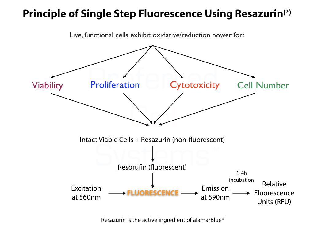
- Incorporates the measurement of non-fluorescent Resazurin (alamarBlue®) to fluorescent Resorufin in an oxidation/reduction reaction that occurs only in live cells. The amount of fluorescence (relative fluorescence units, RFU) is dependent on the metabolic status of the cells and therefore is not only a measure of metabolic viability, but cell proliferation or inhibition.
- After culture, add 10μL of reagent, mix and measure signal between 30 minutes and 4 hours in a fluorescence plate reader.
- Uses a plate fluorometer or multi mode plate reader to measure fluorescence with an excitation filter 560nm and an emission filter of 590nm.
For Research Use Only. Not for clinical diagnostic use.
Fluorescence or multimode plate reader with a 560nm excitation and 590nm emission filter.
- AlamarBlue™/Resazurin Reagent
- 3% SDS solution to stop the reaction.
- Resorufin to standardize the assay.
- Non-sterile, black, 96-well plates for assay standardization.
- Sterile, black, 96-well plates, for cardiac myocyte culture. Choice of adherent or non-adherent plates.
- Sterile, adhesive foil covers to maintain sterility of unused wells.